Hosted by
BioNexus KC
About this event
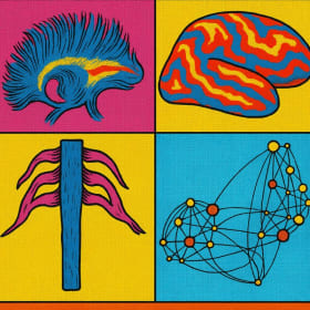
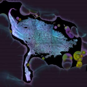
NeuroPop
$450
Starting bid
12x12, Black Frame
Avner Meoded, Children's Mercy
This vibrant pop art image showcases different parts of the nervous system. both central (like the brain and spinal cord) and peripheral (such as the nerves branching out from the spine), using colorful, Pop art-style illustrations.
Each quadrant highlights a unique view created from advanced MRI scans, transformed into an artistic interpretation that makes the complexity of the brain and nerves easier to appreciate. The goal of the image is to spark curiosity and empower kids and families to learn how the nervous system connects and communicates throughout the body.

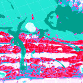
Fireflies
$400
Starting bid
10x10, Natural Frame
Hope Keane, Kansas City University
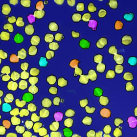
This image was created using bacterial streaking plates. In the lower right corner, you can see the original, unedited lineup of these bacterial plates used to build the final image. The coloration was accomplished through false coloring using our gel electrophoresis imaging machine.
This research is used to establish a procedure to genetically modify diatoms to create a silica-based material to detect various environmental pathogens.
Muscle Terrain
$500
Starting bid
18x13, White Frame
Ziwei Zhao, Kansas State University
This image captures the intricate structure of muscle tissue from the fruit fly (Drosophila), a widely used model in genetic research. The long, white-striped structures represent muscle fibers, responsible for generating movement. Mitochondria, shown in red, are the cell’s energy producers, supplying the power muscles need to contract. A tree-like structure in cyan represents the trachea. The white and cyan circle overlay highlights amyloid structure in muscle.
This study focuses on a genetic muscle condition involving the protein CryAB, which is required to keep other proteins properly folded within muscle cells. In diseased states, CryAB misfolds and aggregates into amyloid-like vesicles that collect and may even leak out of muscle cells, spreading the infection. Understanding how these protein clumps form and spread will aid researchers in developing treatments for muscle degeneration and related hereditary illnesses.

Iridescent Ischemia
$550
Starting bid
16x16, Black Frame
Ava Fleury, University of Missouri
This image captures medical research using a high-resolution laser microscope to study calcium activity in heart cells.
Calcium is essential for each heartbeat. In a healthy heart, calcium moves in a rhythmic cycle within cells, ensuring a steady rhythm. But during events like a heart attack, calcium flow becomes erratic and uncoordinated.
In this image, a mouse heart underwent ischemia—reduced oxygen similar to what occurs in a heart attack. The disrupted calcium signals, no longer synchronized in space or time, were captured using color to represent different phases of the ischemic event. Each heart cell paints a unique hue on the canvas, blending scientific insight with artistic expression to visually depict the chaos of a distressed heart.

Microbe Art
$400
Starting bid
10x10, White Frame
Tamara Cessna, North Kansas City Hospital
Agar art involves the careful cultivation of bacteria, fungi and other microbes on nutrient-rich agar, a gelatinous substance used to grow and differentiate microorganisms in laboratories. Microbiologist 'artists' use different microbial strains, each producing unique pigments, to "paint" on agar plates into a design. Over time, these microbes grow and develop, revealing colorful, living masterpieces.
In this photo, the colors and textures of the flower on the agar art come from the natural pigments produced by microorganisms, including strains of Citrobacter, Pseudomonas, Bacillis, Serratia, Salmonella and Shigella.
Artistic scientists carefully select the microbes (using quality control organisms) to craft designs, accounting for factors such as growth rate, pigment intensity and interaction with other organisms.
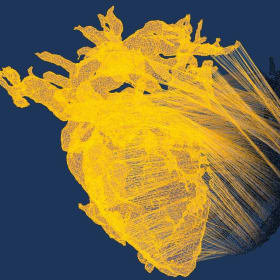
Elastic Heart
$450
Starting bid
16x11, White Frame
Christian Masters, Children's Mercy
This image was created during the preparation of a 3D heart model intended for surgical planning. While 3D imaging is invaluable for pre-surgical visualization, the physicality of a printed model offers surgeons a more tangible sense of scale and spatial relationships, critical for precise, effective procedures. This image is actually heart model editing error.
From Root to Radiance
$650
Starting bid
20x24, Black Frame
Garrett Kyser, Ronawk
This is a layered fluorescent image (Z-stack) of a sheet of HEK-293 cells grown on a hydrogel that mimics human tissue. HEK-293 cells are usually grown in bioreactors to develop biologics for various therapeutic techniques.
The image title reflects how research focused on mass-producing biologics, often without attention to visual structure, can reveal unexpected beauty when the cells are grown on the BioBlock™.
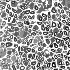
Pathways of Perception
$450
Starting bid
20x15, Black Frame
Javier Llorente Torres, University of Missouri
This image shows the intricate architecture of the peripheral nervous system. Each dark ring represents a single nerve fiber wrapped in a protective layer called myelin, which helps electrical signals travel quickly from the spinal cord to the muscles. The pattern and size of these rings can tell us a lot about nerve health.
This research is focused on a rare genetic disease called SMARD1 (Spinal Muscular Atrophy with Respiratory Distress Type 1), which causes nerve degeneration and muscle weakness in infants. One goal of this research is to understand how the disease damages nerves and how potential treatments might protect or restore them.
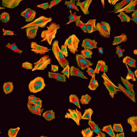
Elegy for a Wandering Cell
$550
Starting bid
14x20, Black Frame
Piyanka Hettiarachchi, Children's Mercy
This image shows glioblastoma cells, a type of aggressive brain cancer, stained to highlight key parts involved in how these cells move and spread. The actin filaments, which are part of the cell’s inner framework, are shown to reveal the structures that help the cells shift and invade surrounding tissue. The nuclei, or centers of the cells, are also marked.
The image was taken four days after a gene-silencing treatment meant to affect cell movement.
Landscape
$450
Starting bid
14x8, Black Frame
Ryszard Jankowiak, Kansas State University
The image shows a high-resolution slice of a 3D distribution of exciton lifetimes in one of the minor light harvesting antennas of Photosystem II from spinach. light-harvesting complexes play a crucial role in photosynthesis by capturing and channeling light energy to reaction centers where it can be converted into chemical energy.
Pollination Odyssey
$500
Starting bid
16x13, White Frame
Brady Blede, Ronawk
This image shows the pollen spores of a White Pine (Pinus strobus) collected in May 2025 in Overland Park, Kansas. The pollen spores appear boat-shaped with two air sacs, called sacci, which give the pollen a winged appearance, helping the spore travel long distances in the wind.
The sample was imaged using light microscopy.
Culturing Saints
$550
Starting bid
24x16, Natural Frame
Emerson Fajardo, Ronawk
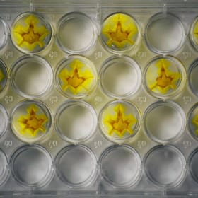
Ronawk's Bio-Block is a 3D hydrogel that allows cells to form tissues as they would naturally. This technology enables rapid expansion of stem cells that could be used for early-stage diagnostics or in repairing injured tissues. These Bio-Blocks had been left to dry, and developed into this Fleur-de-Lis, a symbol connected to New Orleans Saints. Which effectively act like saints with the help they can provide to people.
Blossoms of AlphaFold
$650
Starting bid
24x14, Black Frame
Stefanie Williams, Stowers Institute for Medical Research
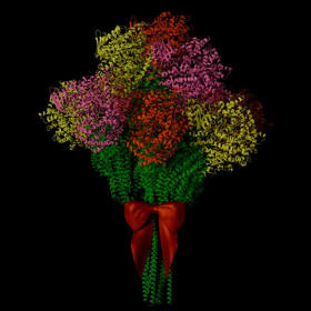
This artwork was created using an open source software called Blender to replicate a predicted protein structure multiple times. The original structure was generated by AlphaFold, a deep learning tool that models how proteins fold and interact. In this case, the interaction between two proteins from the artist’s research unexpectedly resembled a flower, complete with stem and leaves.
Each flower-like structure was colored differently and arranged into a bouquet. An open source bow was added as a final touch. The piece highlights how scientific tools can inspire creativity and transform complex data into visual beauty.

Denatured Chromosome
$350
Starting bid
16x20, Canvas (original)
Michay Diez, Stowers Institute for Medical Research
This original painting depicts a denatured chromosome, one that has lost its usual structure. In its unraveled state, the image reflects both the hidden mysteries within our genetic code and the potential it holds for solving current and future challenges in human disease.

Deeper into the Mind
$450
Starting bid
18x13.5, Natural Frame
Shreya Katwala, University of Missouri-Kansas City
This image represents research into how high oxygen levels affect brain development, particularly in newborns exposed to medical oxygen. It focuses on the cerebellum, the part of the brain responsible for motor control and coordination.
In conditions such as bronchopulmonary dysplasia (BPD), a lung disease common in premature infants, prolonged exposure to high oxygen can lead to inflammation in both the lungs and the brain. This image shows a mouse cerebellum stained for IBA1, an inflammatory marker. The darker dots indicate areas of active inflammation, revealing potential sites of damage caused by excessive oxygen, a condition known as hyperoxia. This signals ongoing injury to the brain.
Understanding how oxygen therapy impacts the developing brain is critical to improving long-term outcomes for infants born prematurely.
Did you know? We fundraise with Zeffy to ensure 100% of your purchase goes to our mission!